The case of a skin lesion on the wrist.
Clinical on-line discussion and reflection. Part-2
seborrhoea keratosis affecting the left wrist in a male (64)
The dermatological presentation of seborrhoeic keratosis is discussed using liquid nitrogen (N2). This article considers the case of a skin lesion on the wrist. The previous article covered differential diagnosis by podiatrists. Material from several sources are explored. This short case demonstrates cryosurgery a on the wrist exemplifying the healing cycle.
Reading score for this article = 55
Seborrhoea keratosis
The following material has been adapted from the British Association of Dermatologists for ConsultingFootPain and Clinician Portal. Last updated October 2017 and due for an update 10/20.
What are seborrhoeic keratoses?
Seborrhoea keratoses are also known as seborrhoeic warts and basal cell papillomas. They are benign growths and very common. They are harmless and often pigmented. In the UK >50% men and >33% women have at least one S. keratosis. By the age of 40, 30% of the population would be affected and by age 70 this increases to 75%. Some people will have only few seborrhoeic keratoses, while others will have large numbers. S.keratoses are not infectious and do not become skin cancerous.
Lesions occur mostly on the trunk, but they are also common on the head and neck. Their numbers vary. One person may have just one seborrhoeic keratosis whilst others can have hundreds. Once present, they usually stay, and new ones often appear over the years.
Aetiology
These lesions have nothing to do with sebaceous glands or viral warts. We don’t know what causes them. It has been suggested that exposure to sunlight and the human papilloma virus (HPV) are risk factors. They are harmless, and usually do not cause symptoms. They can sometimes itch, become inflamed, and catch on clothing.
Presentation
Seborrhoeic keratoses have a rough surface. Their colour can range from golden brown to mid brown to almost black. On dark- skinned people they can appear as multiple small dark brown or black bumps, especially on the face and the neck; in such a case this is called Dermatosis Papulosa Nigra.
Small flat seborrhoeic keratoses can often become raised and larger as the years go by. Their size varies from less than 1 cm to several cms across. They give the impression that they are stuck onto the surface of the skin; however, some look like small pigmented skin tags.
Management of lesions
These lesions are not routinely removed in hospitals. Treatment can occur by either freezing them with liquid nitrogen (cryotherapy), or scraping them off (curettage) under a local anaesthetic. Web link for more details
Reflective Practice
Skin lesions are provoking more clinical comments on-line than ever. The specialty of podo-dermatology is stimulating a new generation of podiatrists to expand their scope of practice and support patients. It has been my view that GP support is important for timely diagnosis.
Cancers and solar damage affecting the skin are more common than we think.
ConsultingFootPain has published the effects of cancer treatment on foot health (Afni Shah-Hamilton). The combined elements of practice associated with skin changes and cancer are attracting considerable interest. by in the past consider the effect of
Differentiating skin lesions
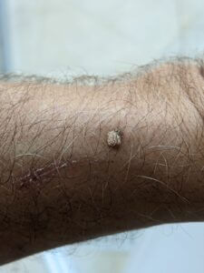
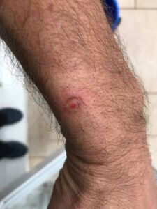
Keratin changes on the surface often have a warty link or appearance. The question arises should we engage with treatment? Raised lesions perhaps occur more on non weight bearing pedal surfaces. In this case, a male (64), the arms were exposed to sun. The appearance of the pigmented base was the reason for the concern. The patient had dorsal trunk lesions of a similar nature. Wide excision would allow a better confirmatory diagnosis based on histology. It would also remove doubts about a MM. Liquid nitrogen was offered and used. Liquid N2 is the mainstay for management.
Liquid nitrogen affected a blister reaction within 8 hours. This appearance lasted for 3 days after the blister appeared.
The effects (photos) of Liquid nitrogen on a lesion
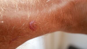
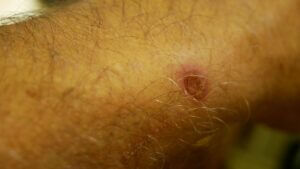
Following Liquid N2 on day 3 the blister burst and the top layer shrunk leaving a dry wound base, open but clean. Most of the peripheral erythema has dissipated.
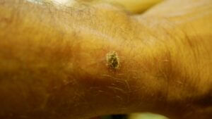
The bulk of the lesion has disappeared at day 4. The right hand picture was taken an hour later after a shower.
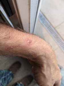
The surface has now left a small crater. A red raised central area is present with young epidermal cells covering the base thinly. The edge of the wound is well demarcated at 4 days post treatment
Day 7 following cryosurgery. The wound is drying up at the centre. The periphery developed a scab. Erythema around the periphery is visible. Active healing is occurring no itching.
Day 18 the wound is healing well and while peripheral erythema is still evident a crusty covering has fully developed while the skin repairs beneath. The dried mass is due to start separating.
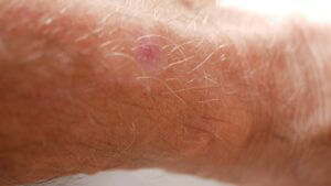
Final picture at 8 weeks – pink lesion after cryosurgery leaves pigmented changes with scar replacement.
Cryotherapy is the standard treatment for small epidermal lesions. It allows for removal without the need for anesthaesia, with no broken skin or bleeding, and with a tolerable amount of pain (taken from Monroe, JR 23/11.W3). This article is worthwhile reading on the effects and benefits together with the indication for using cryosurgery (cryotherapy) published under MDEdge. From the lesion on the wrist (shown) the intentions are to create a process where the blood supply to the basal layers and EDJ are interrupted so as to cause clinical necrosis. The skin cells are then afforded a new chance to regenerate. Monroe, in this 2013 article, makes clear what lesions can and cannot be managed with liquid N2.
The illustrations continue to be updated to complete the series originally posted on UK Podiatry (Facebook) as part of CPD
Thanks for reading ‘Seborrhoeic Keratosis’ written by David R Tollafield for Clinician Portal
Clinician Portal is designed and written for podiatrists and other interested clinicians in managing patients, feet and general health. You can receive a regular newsfeed by signing-up to ConsultingFootPain FREE.
Published by Busypencilcase Reflective Communications
Established 2015
![]()
Published 1 December 2020. Updated 17-01-2021
Recent Comments