
Assessing the Foot with Imaging
A free-view chapter from The Institute of Chiropodists & Podiatrists ‘ – An Introduction to the Foot’.
Imaging is the term for taking specialised pictures of human tissue. Traditionally this was by radiographic X-rays, but refinement to the imaging system has overcome many of the problems first encountered. X-rays use radiation, and large doses were accumulative and caused cancers in their earlier utilisation. Filters and enhancement using special plates today avoid wet film development. Digital pictures are fast and can be manipulated on computers and shared by professionals across the internet. This has sped up the process of diagnosis and action. Anyone requesting X-rays or other imaging in the UK must take an Ionising Radiation (Medical Exposure) Regulations (IRMER) course. While X-rays are still valuable, scanning now has a number of different alternatives. With the exception of ultrasound, most imaging using any X-rays should not be carried out during pregnancy.
X-RAY
X-rays pass through the foot, and bones made of calcium and phosphate absorb radiation while the soft tissue is bypassed. Bone appears white, and the joints where the cartilage lies are dark. Cartilage does not reflect absorption as it is soft tissue. Muscles, ligaments and other soft structures are not observable, although gas from infection is visible.
X-rays, therefore, have optimum value for bone and are used after injury, for suspected fractures, collapsed bone around joints, infection, tumours, and penetrating objects known to provide a signal. Most glass, for example, does not show, while a metal such as a needle or a nail would absorb X-rays. Lead aprons are used to protect clinical staff.
ULTRASOUND (US)
Ultrasound is preferred when soft tissue is predominantly the anatomical part of greatest interest. This uses no radiation and is safe. Diagnostic US is useful for tendons, fascia, joints, and muscles. Ultrasound uses high-frequency sound waves that return an image to the transducer probe. Today, such a diagnostic technique is reliable in experienced and trained hands.
MAGNETIC RESONANCE IMAGING (MRI)
Magnetic Resonance Imaging uses magnets and electricity as the source for this type of image. The image produced is three-dimensional, where the anatomy can be recreated in detail. MRI can be used for soft tissue but also bone. This is safe because no radiation is used, but previous metal work impedes the image quality in the foot and ankle.
MRI uses a doughnut-shaped container. Unlike whole-body scanning, the lower limb can enter the container while the patient stays outside, avoiding any sense of phobia. MRI scanners are noisy, and ear protection is required. The scan can take 30-40 minutes, and it is essential that the patient does not move to avoid blurring.
COMPUTERISED TOMOGRAPHY (CT)
Computerised Tomography scanning offers useful clarity in cross-section and is not dissimilar to MRI in the quality of images. However, the system uses high doses of radiation compared to X-rays, but it does offer an option if metalwork is present. CT can produce constructed high-quality 3D images. CT is better for hard tissue, while MRI is better for softer tissue.
BONE SCAN (NUCLEAR MEDICINE)
In nuclear medical imaging, Technetium99 is attached to another chemical, methylene diphosphonate allowing a chemical attachment with hydroxyapatite in bone. It is useful for picking up areas of bone damaged by injury or with a disease, including cancer. This type of scan does have risks as a radioactive chemical is injected into the body. The chemical is excreted out of the body via the kidneys. It has a short radioactive half-life, with over 90% removed within 24 hours. This type of scan is useful if other investigations fail to reveal inflammation but are less commonly used than other scanning forms.
Clinical Application
The rule of thumb when using imaging is not to use it unless a value can be enacted. Even after the injury, X-rays are not routinely indicated unless the signs clearly indicate a problem. Ultrasound is favoured as a first-line method because it provides the clinician with a safe alternative. Once imaging has been undertaken, the clinician is aided with the best information to design a treatment programme. In a number of cases, fractures in feet are difficult to identify after injury or damage and may take several weeks to show up. Repeating X-rays is sometimes necessary to establish if repair pathology has occurred.

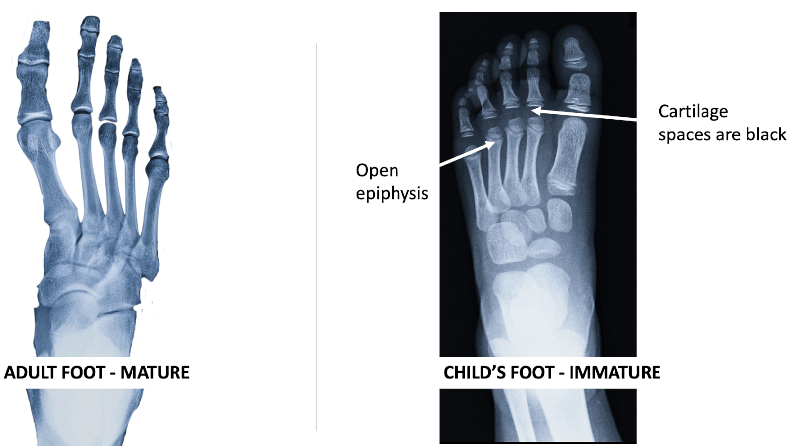
Mature foot versus undeveloped foot pre-adult X-Ray comparison. Click here to see a larger image.
Figure 37: Contrasting developed bone with immature skeletal development. Note that the navicular, cuboid and cuneiforms are underdeveloped, and the sesamoids have not appeared.

BUY NOW
This article has been adapted for ConsultingFootPain from An Introduction to the Foot and Its Common Problems in the Adult.
An Educational publication from the Institute of Chiropodists & Podiatrists for foot health practitioners, nurses and other health professions. ISBN 9781838413736
Available from the Institute of Chiropodists & Podiatrists or directly from Amazon Books
Published by Busypencilcase Communications Est. 2015
![]()
Recent Comments