Does evidence help to explain hammer toe? – Part 2
This is a follow on article to ‘The Puppet Toe‘, published 6th January 2023, which discusses the case of Zandra’s toe problems. Two questions arise. Does the hammer toe come about because of a single factor of influence, or is it hereditary?
In Part 1, Zandra presented with increasing flexion deformity of the index (2nd) and 3rd toes. She had previously had successful bunion surgery yielding complete satisfaction. The tose had deformed over time. The evidence for a single cause is weak, yet some recent literature may help provide a clearer aetiology.
Heredity versus Acquired
Hannan et al. (2015) provided insight into the question of heredity. The study looked at the presence of hallux valgus and lesser toe deformities in 2,446 adults and revealed 1,370 participants with an available link to a family history of common deformity.
“We observed significant moderate to high heritability for hallux valgus (H2 = 0.29–0.89, depending on age and sex) and lesser toe deformity (H2 = 0.49–0.90, depending on age and sex), suggesting that there are genetic variants, common and/or rare, affecting hallux valgus and lesser toe deformity…” The study had problems pairing families and siblings because numbers had to be pooled, reducing the overall cohort.
 Shoe fitting inadequacy is often cited in the acquired toe deformity rather than a hereditary toe. Tight socks or hand-me-down small sizes. Children ideally should be fitted and not use formal shoes too early. My wife was scrupulously careful about her son’s feet, yet he had a bent toe. As he grew into an adult, the problem gave him no trouble and seemed less obvious 40 years later.
When a patient fractures their ankle, the long tendon which runs around the inside bone (malleolus of the tibia) can become scarred and causes a claw toe, as in Tsukamoto, where the authors noted – the “Flexor hallucis longus tendon was stuck together with strong scar tissue, so the scar tissue was removed, improving the toe deformity.” (Tsukamoto, M et al. 2011).
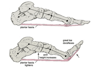
https://www.physio-pedia.com/Windlass_Test is demonstrated
Theory of toe deformity with arch shape
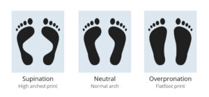
Foot arch variation diagram – supination and overpronation and normally arched human sole footprint drawing
The hindfoot contains the long tendons at the ankle junction inside and outside the joint. Disruption undoubtedly influences tendons distally and may override the short tendons. When the foot flattens or heightens, the muscles’ position and tendons change, exerting a different pull. This concept has been in vogue since the sixties emanating from US podiatry education and scholastic theory. To complicate matters, the involvement of the fascial band cannot be excluded from exerting an effect on the MTP joints. The Windlass mechanism is shown in the schematic above, and the video link shows the foot in motion affecting the arch.
We can learn much about how muscles work by neutralising their effect. The problem lies in the relative difficulty of studying live patients as opposed to those who leave their bodies for science (cadavers). Large muscles have been studied much more effectively because there is a large surface to adhere electrodes to. Of course, dead muscle tissue does not react. Botulinum (Botox) was used in patients with stroke – a condition where the neural pathway from the brain to the muscle is affected, Takekawa, 2022.
In a study conducted as part of the Johnston County Osteoarthritis Project (Golightly, 2014), 1466 subjects with either flat foot (overpronated), neutral or arched (over-supinated) feet were selected to see if the foot type contributed to several key foot problems and deformities. Hallux valgus and lesser toe deformity were included. This study had the advantage of looking at overweight patients and considered a comparison between black (African-American) and Caucasian races. We must remind ourselves that the feet of black races often have a flatter foot in their natural state compared to Caucasians.
N.B. The diagram above provides a broad idea but should not be confused with significant pathologies of pes valgus and, pes cavus, where neuromuscular involvement may be included in the condition. In both conditions, the lesser toes may be adversely affected by the foot position and vector set-up.
Golightly et al.’s (2014) study described the toes as hammer and claw. Zandra had a hammer toe, so the end contacted the ground, and her flexor tendons were tight, having contracted over time. Golightly found 26.7% of his subjects had hammer toes with over-pronation, and 1.8% had claw toes. Of those where the foot was over-supinated, 26.8% were hammer toes, and 1.9% were clawed toes. Those with neither over-pronated nor over-supinated feet had 25.4 and 2.2%, respectively.
Regarding racial difference, the African American foot had a lower incidence of hammer or claw toes for all foot positions and obesity, based on +30 BMI, had no difference compared to underweight subjects. The study implies that hammer toes creep with age and have no gender preference but are racially more prevalent in Caucasians. The study was not supported with dynamic pressure measurement and so, in one sense, cannot be validated.
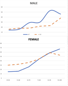
Taken from data from Golightly (2014) comparing HV hallux valgus [broken orange line] with HT hammer toe incidence by age versus gender (see reference)
Pulleys & tendons
We must first realise that foot anatomy is divided into muscles within the foot (intrinsic) and muscles outside the foot that arise in the leg bones (tibia & fibula). These are the extrinsic group. The hands work in the same principle; like a Marionnette, the guides (tendons) control digital movement. The extrinsic tendons are longer, but like the rails of the main train station, they run parallel down the length of the toes. Tendons on top of the foot are called extensor tendons and lift the toe (extend). Those lying underneath are flexor tendons and cause the toe to bend. The toe adopts that hammer-like position when the toe bends too much or stays bent. The smaller muscles, such as the lumbricals, modify the tendon pull, acting through the extensor sling mechanism.
A Toe flexion pattern was observed during electrical stimulation of two tendons influencing flexion – the long, big toe tendon to the first toe (FHL) and the long, smaller toe tendon (FDL). The stroke patients had claw-foot deformities treated with Botox. The FHL and FDL tendon arrangement was also studied in five limbs of three cadavers. The toe flexor tendon will pull if the muscle is stimulated with an electrical impulse. The big toe flexed in all cases, but the second toe responded in 25 out of 28.
Cadaver studies showed the division of the FHL tendon with branches fusing with the FDL tendon in all five limbs examined; none of the tendons was inserted only in the first toe, (Takekawa, T. et al. 2022). The same authors stated that “movements of the second and third toes are controlled by both the FDL and FHL muscles. The findings highlight the need for BoNT (Botox) injection in both the FDL and FHL muscles for the treatment of claw-toe deformity.”
How does the body stop toes bending?
If we think back to physics, a pulley provides mechanical advantages for moving a heavy object. The pulley, in effect, changes the direction of the force. In foot biomechanics, G. James Sammarco (1982) described the effect of the relationship between the intrinsic and extrinsic tendons via a sling mechanism (see a diagram of a finger). The sling mechanism is still a pulley (extensor hood) and prevents overpulling of the stronger flexor tendons.
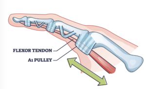
This is purely schematic rather than an accurate representation of the toe. An extensor band lies around the foot MTP joints (not shown – Frankel & Nordin). This picture shows a finger, not a toe, to illustrate the annular bands.
Taffur (2015) used cadaver anatomy to identify pulleys – as shown in the diagram. Fibre rings exist as supplementary pulleys made up of fibres of a protein called collagen and are arranged in rings. Taffur showed the ring arrangement using high-performance magnetic resonance (MR) scanning – “it would have been unlikely that it would be seen with routine clinical systems. Exquisitely detailed anatomy of the pulley system of the flexor tendons of the toes was apparent… These features had not been described previously in MR studies of the finger pulleys as well as similarities and differences between the pulley systems of the toes and fingers.”
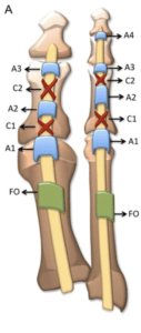
Taffur (2015) see references the first & second toes from the underneath aspect.
The A2 pulley in the great and lesser toes and the A4 pulley in the lesser toes were the most substantial. The A5 pulley, which was not previously described in the toes, was demonstrated. The cruciform pulleys were also seen and were smaller and thinner. Three tissue layers were seen, and there was evidence of different fibre directions in annular pulleys producing different magic angle effects.
Diagrammatic representation of the pulley system of the flexor tendons in the foot a and the hand b in accord with the literature. The A5 and C3 pulleys are absent in the lesser toes compared to the finger. The localization of the A4, C1 and C2 pulleys in the lesser toes significantly differs from the fingers and can be appreciated in the illustrations. There is one additional pulley (A3 pulley) in the great toe. The thumb lacks cruciform pulleys and has an oblique pulley (OP in B) opposite the proximal phalanx. The A2 pulley also has a different location in this two-pulley system. FO, Fibrous oblique band [vi]
Conclusion
This article commenced with Zandra and her toe deformities in Part 1. The cause balances an argument based on a natural hereditary cause and an imbalance in the mechanics. The evidence of a dysfunctional hallux valgus, stiffened through surgery, provides another tangible cause of her problem, as the FDL is associated with the function of the FHL. However, Zandra’s right foot was operated on simultaneously and has not revealed the same toe deformities. In this foot, her toes are well aligned. The second toe is a major foot stabilising force next to the great toe. It also has a longer length as a general rule than the lateral 3rd – 5th toes. Knock out the first toe, so it does not have the flexible function, and a second toe overpull could well arise. The evidence emerging is that toe deformities can creep up on male and female patients with advancing age. Some hallux valgus corrections do less well than others, and secondary features may or may not be associated with such failure. While that failure is far from an accusation of negligence, it should be recognised by all specialists dealing with the foot who offer surgical advice.
The evidence gathered here is inconclusive because research seldom can create a scenario covering all aspects of foot function. As technology improves, we can research smaller muscle functions methodically at the clinical level. For the time being, we rely on smaller projects to build a picture.
- Hannan, M.T., Menz, H.B., Jordan, J.M., Cupples, L.A., Cheng, C.-H. and Hsu, Y.-H. (2013), High Heritability of Hallux Valgus and Lesser Toe Deformities in Adult Men and Women. Arthritis Care & Research, 65: 1515-1521.
- Tsukamoto, M., Furuichi, I., Murata, M., Miyata, N., Moriguchi, N., Yoda, I., & Eto, S. (2011). Claw toe deformity after ankle fracture: A case report. Seikeigeka to Saigai Geka, 60(1), 112-116. https://doi.org/10.5035/nishiseisai.60.112[NB – this paper is in Japanese if downloaded]
- Golightly YM, Hannan MT, Dufour AB, Hillstrom HJ, Jordan JM. Foot Disorders Associated With Overpronated and Oversupinated Foot Function: The Johnston County Osteoarthritis Project. Foot & Ankle International. 2014;35(11):1159-1165. doi:10.1177/1071100714543907
- Takekawa, T., Takagi, S., Kitajima, T., Sato, T., Kinoshita, K., & Abo, M. (2022). Claw Toe: Anatomic Guide for Injection of Botulinum Toxin into Foot Muscles. Canadian Journal of Neurological Sciences / Journal Canadien Des Sciences Neurologiques, 49(1), 102-108. doi:10.1017/cjn.2021.52
- Sammarco, GJ. The foot and ankle in classical ballet and modern dance. In Disorders of the foot. Vol.2. Edited MH Jahss. Philadelphia. WB Sanders. pp1629-1659
- Tafur, M., Iwasaki, K., Statum, S. et al. Magnetic resonance imaging of the pulleys of the flexor tendons of the toes at 11.7 T. Skeletal Radiol 44, 87–95 (2015).
Reference for Professionals
- Beldame, J., Lalevée, M., Regnard, S. et al. Impact of intertendinous connections between the flexor digitorum brevis and longus on percutaneous tenotomy for the treatment of claw toes: an anatomic and ultrasound study. Surg Radiol Anat 43, 1067–1073 (2021). https://doi-org.libaccess.hud.ac.uk/10.1007/s00276-021-02723-8
- Frankel, VH, Nordin, M. Basic Biomechanics of the Musculoskeletal System. Second edition. Lea and Febiger. 1980. pp 172-174
Thanks for reading ‘Thoughts on the Hammer Toe’ by David R Tollafield
![]()
Published by Busypencilcase Communications Est. 2015 for ConsultingFootPain

Recent Comments